
Your cart is currently empty.
Saving an immature tooth with a deep vertical fracture
Dive into a highly complex case involving an immature fractured tooth.
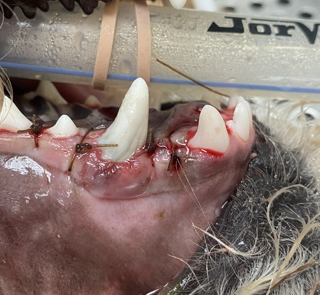
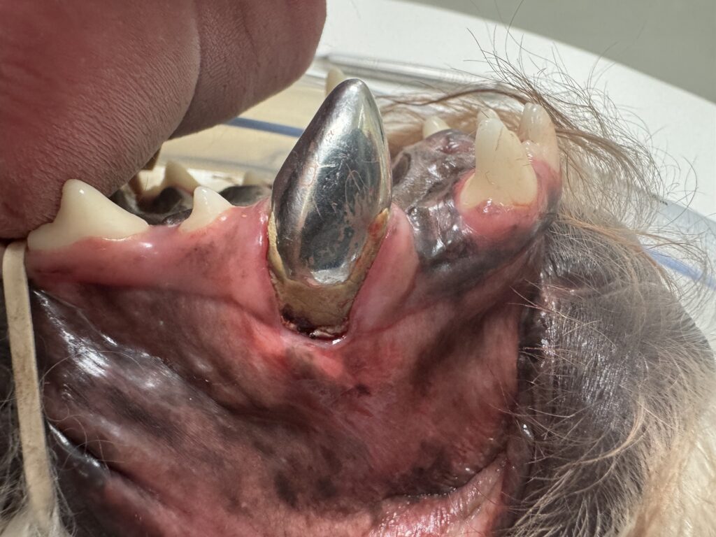
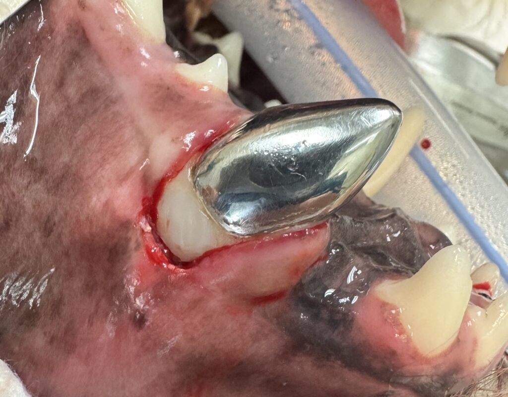
A 6.5-month-old standard poodle was presented to VDS for a fracture of the maxillary left canine (204). (Figures 1a-1c)
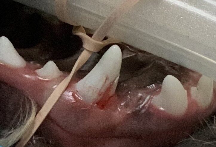
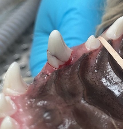
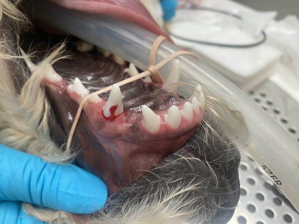
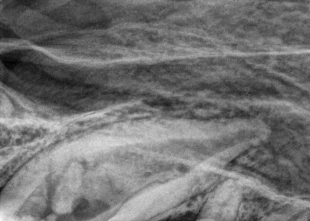
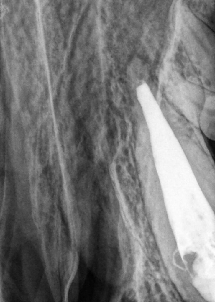
Normally, fractured teeth can have a very good prognosis; however, two issues complicated the procedure in this case. First, there was a deep subgingival fracture on the tooth. This extended below the bone, which will create periodontal issues. In addition, because the patient was so young, the root was not complete (Figure 2), and thus standard root canal therapy was not an option.
To save this tooth, multiple procedures would be required, and each would have a questionable prognosis. The combination of these issues made the overall prognosis for a successful, long-term, infection-free maintenance of this tooth poor. All of this was discussed with the owner, and extraction was recommended; however, due to his age, they elected to proceed with the process of salvaging the tooth.
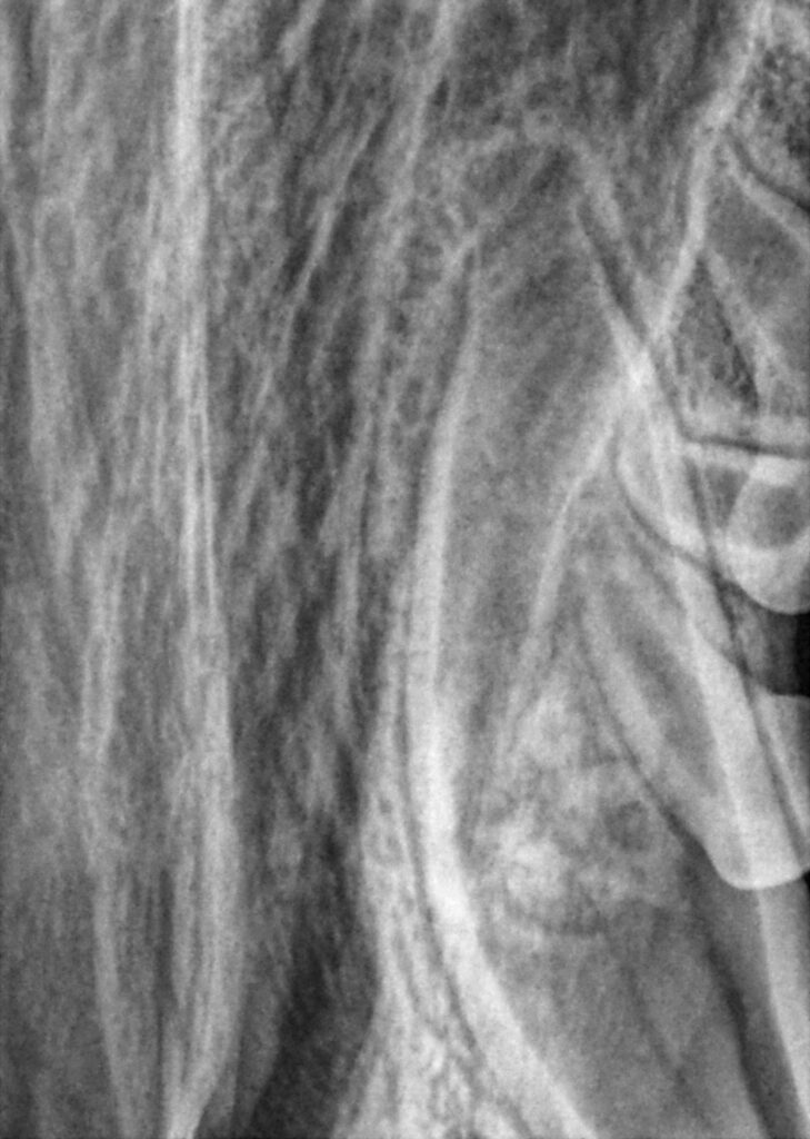
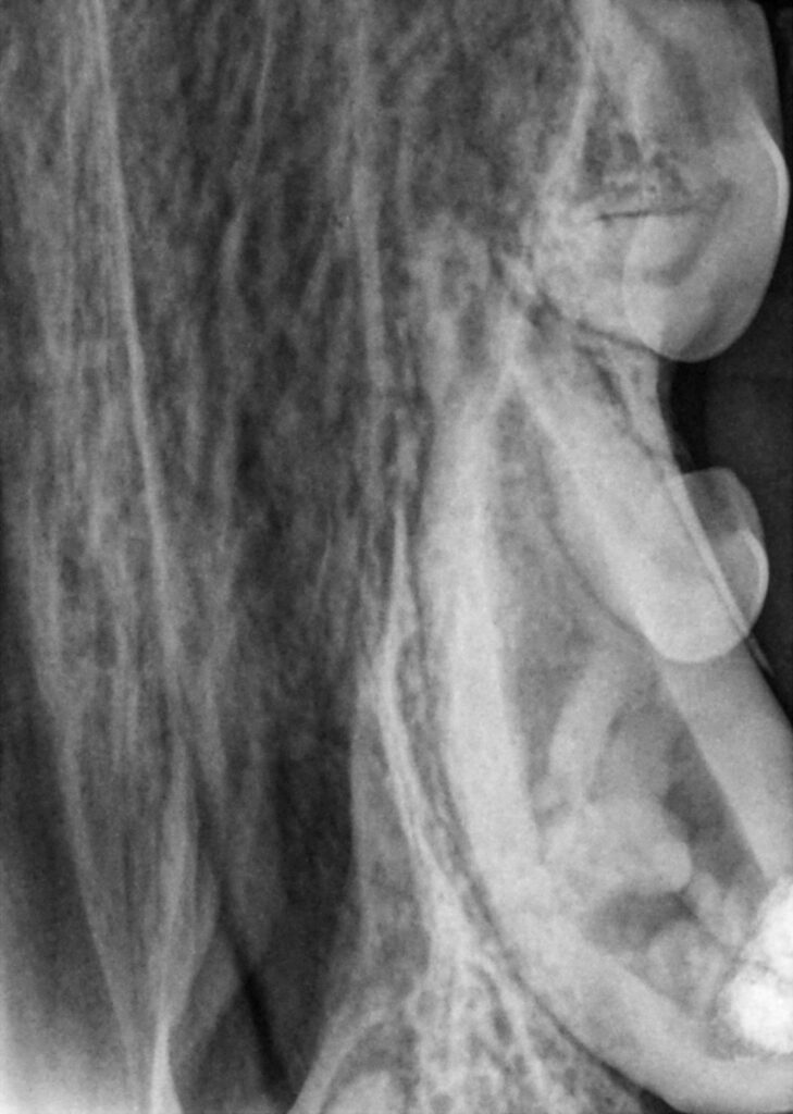
The patient was placed under general anaesthesia, and an exam and dental radiographs were performed (Figure 3). This confirmed the absence of a complete apex and the vertical root fracture.

The first step was to create a periodontal flap to restore the vertical root fracture and prevent leakage. Next, vital pulp therapy was performed (Figure 3) to protect the tooth and (hopefully) allow it to mature to the point of receiving root canal therapy in the future. The subgingival defect was restored with glass ionomer to allow for superior periodontal attachment. Finally, the flap was sutured (Figure 4).
The patient was re-presented 9 months later for follow-up (6 months was recommended). Per the owner, the tooth had broken 2 weeks previously. Upon presentation, the tooth was found to be further broken, with about ½ of the tooth missing and the subgingival restoration lost (Figures 5a and 5b). The tooth was necrotic.

The patient was placed under general anaesthesia and an exam and dental radiographs exposed. The images revealed that the tooth had lived long enough to complete apexogenisis; however, there was slight periapical lucency, indicating endodontic infection (Figure 6a and 6b).
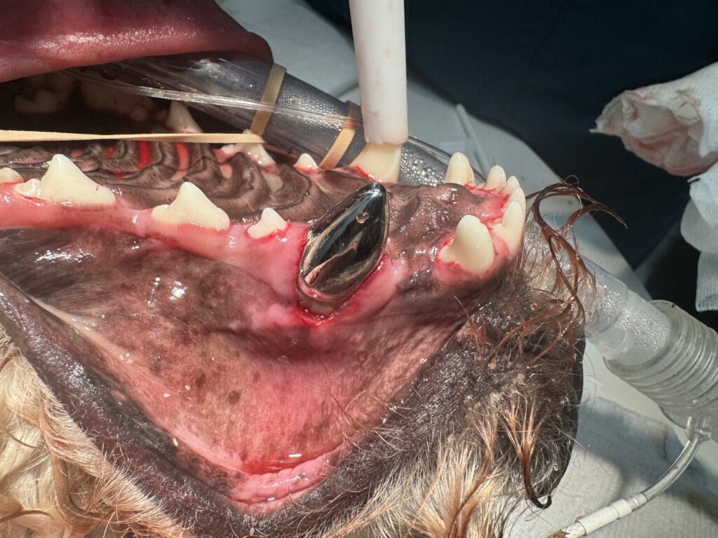
The tooth was treated with standard endodontic therapy (Figure 7) and prepared for a titanium crown (Figure 8). The client was instructed on home care and to recheck in 6 months.

One year later, the pet was presented for follow-up. The client had not been very compliant with home care. On presentation, the crown was intact with mild to moderate gingival recession, but minimal inflammation (Figure 9). Dental radiographs were exposed, which revealed that the root canal was successful and there was minimal to no progression of periodontal loss. (Figure 10). The tooth was cleaned, and the client was instructed on home care.
This case represents a successful result of a highly complex case requiring two endodontic treatments, periodontal surgery, and multiple advanced restorative procedures. Despite the poor prognosis (and client compliance), the tooth was saved and has a decent long-term prognosis.
Learn more about broken or fractured teeth.